USG-Guided FNAC
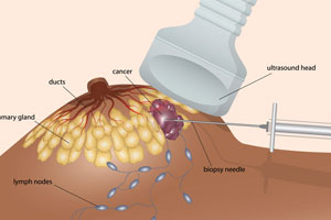
USG-Guided FNAC
USG-Guided FNAC (Ultrasound-Guided Fine Needle Aspiration Cytology) is a minimally invasive diagnostic procedure that uses ultrasound guidance to accurately collect cells from a lump, swelling, nodule, or abnormal area for microscopic examination. It helps diagnose various conditions affecting the thyroid, lymph nodes, breast, salivary glands, soft tissues, and other superficial organs with excellent precision and safety.
At Dr. Dere Sonography & X-Ray Centre, USG-Guided FNAC procedures are performed by Dr. Sameer Dere, Consultant Radiologist, using advanced ultrasound guidance techniques to ensure accurate needle placement, proper sample collection, patient comfort, and reliable diagnostic results.
Unlike conventional FNAC performed without imaging guidance, USG-Guided FNAC allows real-time visualization of the target lesion. This improves accuracy by helping the radiologist precisely guide the needle into the area of concern while avoiding surrounding important structures.
USG-Guided FNAC is commonly recommended for evaluating thyroid nodules, breast lumps, enlarged lymph nodes, and other suspicious lesions. The procedure provides valuable diagnostic information that helps doctors determine the appropriate treatment approach.
Conditions Evaluated During USG-Guided FNAC
USG-Guided FNAC helps obtain tissue samples from various body regions for accurate diagnosis of multiple conditions.
- Thyroid nodules and thyroid swellings.
- Breast lumps and breast lesions.
- Enlarged lymph nodes.
- Salivary gland lesions.
- Soft tissue swellings.
- Cysts and fluid-filled lesions.
- Subcutaneous masses.
- Suspicious nodules requiring cytological evaluation.
- Benign and malignant lesion assessment.
- Follow-up evaluation of previously diagnosed lesions.
- Other superficial lesions as advised by the treating physician.
When is USG-Guided FNAC Recommended?
Your doctor may recommend USG-Guided FNAC whenever a detailed cellular evaluation of a lump, nodule, or abnormal tissue area is required for diagnosis and treatment planning.
- Presence of a thyroid swelling or thyroid nodule.
- Evaluation of breast lumps or suspicious breast lesions.
- Persistent or enlarged lymph nodes.
- Unexplained soft tissue swellings.
- Assessment of lesions detected on ultrasound imaging.
- Need for cytological confirmation before treatment.
- Monitoring previously diagnosed lesions.
- Whenever advised by your surgeon, physician, endocrinologist, or oncologist.
Why is USG-Guided FNAC Recommended?
USG-Guided FNAC is recommended when accurate cellular evaluation of a lump, swelling, or abnormal lesion is required. Ultrasound guidance allows precise needle placement, improving sample accuracy while reducing the risk of injury to surrounding structures.
- Provides accurate sampling from suspicious lesions.
- Allows real-time ultrasound guidance during the procedure.
- Improves diagnostic accuracy compared to blind FNAC.
- Helps differentiate benign and suspicious lesions.
- Supports early diagnosis and treatment planning.
- Minimally invasive procedure with minimal discomfort.
- Useful for evaluating lesions in difficult-to-access locations.
- Provides valuable information for surgeons, physicians, and oncologists.
What Does USG-Guided FNAC Assess?
USG-Guided FNAC helps obtain cell samples from targeted lesions under continuous ultrasound visualization. It allows accurate assessment of various swellings and nodules to determine their nature and guide further management.
Thyroid Nodule Assessment
USG-Guided FNAC is commonly performed for thyroid nodules to evaluate cellular characteristics and help determine whether a nodule is benign or requires further investigation.
Breast Lesion Evaluation
The procedure helps assess breast lumps and suspicious lesions by obtaining cell samples for microscopic examination and accurate diagnosis.
Lymph Node Evaluation
Enlarged or abnormal lymph nodes can be evaluated through targeted FNAC to identify infections, inflammatory conditions, or other pathological changes.
Soft Tissue Lesion Assessment
USG guidance helps accurately sample superficial soft tissue swellings, cysts, and masses for diagnostic evaluation.
Salivary Gland Lesion Evaluation
The procedure assists in evaluating salivary gland swellings and abnormalities by obtaining cells from the affected area with precise ultrasound guidance.
Cystic Lesion Assessment
Fluid-filled lesions and cysts can be evaluated to determine their nature and assist doctors in planning appropriate management.
Suspicious Mass Evaluation
Any suspicious superficial mass detected clinically or on ultrasound can be accurately sampled to help establish a diagnosis.
Follow-up Lesion Assessment
Previously diagnosed lesions can be re-evaluated when changes in size, appearance, or clinical symptoms require further investigation.
How is USG-Guided FNAC Performed?
USG-Guided FNAC is a minimally invasive procedure performed using ultrasound imaging to guide a fine needle accurately into the target lesion. The procedure usually takes around 15 to 30 minutes and does not require major preparation or hospitalization.
- Your medical history, ultrasound findings, and previous reports are reviewed before the procedure.
- The targeted area is identified using high-resolution ultrasound imaging.
- The skin over the area is cleaned using sterile precautions.
- A fine needle is gently guided into the lesion under continuous ultrasound visualization.
- Cell samples are collected and sent for cytological examination.
- A detailed procedure report is prepared for your treating doctor.
Benefits of USG-Guided FNAC
USG-Guided FNAC combines the accuracy of ultrasound imaging with minimally invasive sampling, making it a valuable diagnostic procedure for evaluating various lesions and swellings.
- Precise needle placement using real-time ultrasound guidance.
- Improved sample accuracy for reliable diagnosis.
- Minimally invasive procedure with minimal discomfort.
- No surgical incision required.
- Quick procedure with faster recovery.
- Helps detect abnormalities at an early stage.
- Safe evaluation of thyroid, breast, lymph nodes, and soft tissue lesions.
- Supports accurate treatment planning through cytological diagnosis.
Preparation Before USG-Guided FNAC
USG-Guided FNAC is a simple, safe, and minimally invasive diagnostic procedure that requires minimal preparation. Following your doctor's instructions and carrying relevant medical records helps ensure accurate evaluation and smooth completion of the procedure.
- Carry your doctor's prescription along with previous ultrasound reports, FNAC reports, biopsy reports, or relevant medical records.
- Inform your doctor about any blood-thinning medications or bleeding disorders before the procedure.
- Wear comfortable clothing that allows easy access to the area being examined.
- No fasting is usually required unless specifically advised by your doctor.
- Inform the radiologist about any allergies, previous procedures, or medical conditions.
- Arrive at the diagnostic centre 10 to 15 minutes before your scheduled appointment.
Is USG-Guided FNAC Safe?
Yes. USG-Guided FNAC is a safe, minimally invasive, and commonly performed diagnostic procedure. Since the needle is guided using real-time ultrasound imaging, the procedure allows accurate sampling while minimizing injury to surrounding tissues. It usually involves minimal discomfort and does not require hospitalization.
At Dr. Dere Sonography & X-Ray Centre, USG-Guided FNAC procedures are performed using advanced ultrasound guidance techniques while maintaining strict hygiene standards, patient safety, comfort, and diagnostic accuracy.
Frequently Asked Questions
What is USG-Guided FNAC?
USG-Guided FNAC is a diagnostic procedure where ultrasound imaging is used to guide a fine needle into a lump, nodule, or abnormal area to collect cells for microscopic examination.
Is USG-Guided FNAC painful?
The procedure is generally well tolerated. Most patients experience only mild discomfort or a small pinching sensation during needle insertion.
How long does USG-Guided FNAC take?
The procedure usually takes around 15 to 30 minutes, depending on the location and nature of the lesion being evaluated.
Will I need admission after FNAC?
No. USG-Guided FNAC is an outpatient procedure, and most patients can return home shortly after completion.
When will I receive the FNAC report?
The collected sample is examined by a cytopathologist, and the reporting time depends on the type of examination required and laboratory processing.
Why Choose Dr. Sameer Dere for USG-Guided FNAC?
- Consultant Radiologist with expertise in Ultrasound-Guided Diagnostic Procedures.
- Advanced ultrasound technology for precise lesion localization and needle guidance.
- Accurate evaluation of thyroid, breast, lymph nodes, and soft tissue lesions.
- Real-time ultrasound guidance for safe and accurate sample collection.
- Detailed procedure documentation and reporting for better clinical decisions.
- Patient-friendly approach focused on comfort and safety.
- Strict hygiene and infection-control practices during the procedure.
- Trusted diagnostic services focused on accurate diagnosis and quality patient care.
Accurate Diagnosis Through Advanced Ultrasound Guidance
Early and accurate diagnosis plays an important role in effective treatment planning. USG-Guided FNAC provides a safe and precise method for evaluating lumps, nodules, and abnormal lesions by combining real-time ultrasound imaging with targeted cellular sampling.
Schedule your USG-Guided FNAC with Dr. Sameer Dere at Dr. Dere Sonography & X-Ray Centre for advanced ultrasound-guided diagnosis, expert evaluation, and compassionate patient care using modern imaging technology.