Neurosonogram
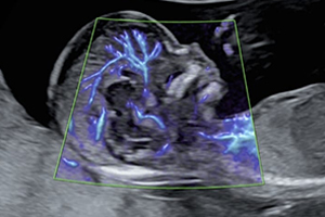
Neurosonogram
Neurosonogram is a specialized ultrasound examination used to evaluate the brain and nervous system structures, especially in newborns and infants through the natural opening called the fontanelle. It provides detailed assessment of brain development, fluid spaces, and possible abnormalities in a safe and radiation-free manner.
At Dr. Dere Sonography & X-Ray Centre, Neurosonogram examinations are performed by Dr. Prajakta Samir Dere, Consultant Radiologist with expertise in Obstetric, Fetal & Gynecological Imaging and pediatric imaging evaluation. Advanced ultrasound technology is used to provide accurate assessment while ensuring patient comfort and safety.
Neurosonogram is commonly performed in newborn babies and infants where the skull bones are not completely fused, allowing ultrasound waves to pass through the fontanelle and provide clear images of the developing brain.
This examination helps evaluate brain structures, ventricles, fluid spaces, and blood flow patterns. It plays an important role in detecting certain neurological conditions early and supports timely medical management.
Conditions Evaluated During Neurosonogram
Neurosonogram helps assess the developing brain and detect various neurological abnormalities in newborns and infants.
- Brain development assessment in newborns.
- Ventricular system evaluation.
- Hydrocephalus (excess fluid accumulation in the brain).
- Intracranial bleeding assessment.
- Brain cyst evaluation.
- Structural brain abnormalities.
- Periventricular changes in premature babies.
- Assessment of brain fluid spaces.
- Follow-up monitoring of known neurological conditions.
- Overall brain health evaluation in infants.
When is Neurosonogram Recommended?
Your doctor may recommend Neurosonogram when detailed evaluation of the baby's brain and neurological structures is required due to clinical concerns, risk factors, or abnormal findings.
- Premature birth or low birth weight babies.
- Abnormal head growth patterns.
- Suspected brain abnormalities.
- Seizures or abnormal neurological symptoms.
- History of birth-related complications.
- Monitoring high-risk newborns.
- Follow-up evaluation of previously detected abnormalities.
- Whenever advised by a pediatrician or neonatologist.
Why is Neurosonogram Recommended?
Neurosonogram is recommended when detailed evaluation of the baby's brain and neurological structures is required. It provides valuable information about brain development, fluid spaces, and possible abnormalities while being a safe and radiation-free imaging technique.
- Provides detailed assessment of newborn brain structures.
- Helps detect brain abnormalities at an early stage.
- Evaluates brain ventricles and fluid spaces.
- Helps identify bleeding or structural changes inside the brain.
- Useful for monitoring premature and high-risk infants.
- Allows follow-up assessment of neurological conditions.
- Safe and painless examination without radiation exposure.
- Supports early diagnosis and appropriate treatment planning.
What Does Neurosonogram Assess?
Neurosonogram provides detailed imaging of the developing brain in newborns and infants. It helps evaluate brain structures, fluid spaces, and possible abnormalities through ultrasound technology.
Brain Structure Evaluation
Neurosonogram helps visualize important brain structures and assess their development, appearance, and overall condition.
Ventricular System Assessment
The examination evaluates the brain ventricles and helps identify enlargement or abnormalities related to cerebrospinal fluid circulation.
Hydrocephalus Evaluation
Neurosonogram helps detect excess fluid accumulation within the brain spaces and supports monitoring of hydrocephalus.
Intracranial Bleeding Assessment
The scan helps identify bleeding within the brain, especially in premature or high-risk newborns.
Brain Cyst Evaluation
Neurosonogram can help detect and monitor certain cystic changes within the brain structures.
Premature Baby Brain Monitoring
Premature infants may require neurosonogram evaluation to monitor brain development and detect possible complications.
Blood Flow Assessment
Doppler techniques may be used when required to evaluate blood flow patterns within brain-related structures.
Follow-up Neurological Evaluation
Neurosonogram can be repeated when required to monitor previously detected abnormalities and assess changes over time.
How is Neurosonogram Performed?
Neurosonogram is a safe and painless ultrasound examination performed through the baby's fontanelle (soft spot on the head). The procedure usually takes around 15 to 30 minutes, depending on the assessment required and the baby's condition.
- The baby's medical history and previous reports are reviewed before the examination.
- The baby is positioned comfortably for the scan.
- Ultrasound gel is applied over the fontanelle area.
- A small ultrasound probe is gently placed over the soft spot of the baby's head.
- Images of brain structures and fluid spaces are obtained.
- A detailed report is prepared based on the findings.
Benefits of Neurosonogram
Neurosonogram is an important diagnostic tool for evaluating infant brain health while ensuring safety and comfort.
- Safe and radiation-free imaging technique.
- Painless and non-invasive examination.
- Early detection of brain abnormalities.
- Useful for premature and high-risk newborns.
- Helps monitor neurological development.
- Allows repeated follow-up scans when required.
- Provides valuable information for pediatric care planning.
- Comfortable examination for newborns and infants.
Preparation Before Neurosonogram
Neurosonogram is a simple, safe, and painless ultrasound examination that requires minimal preparation. Proper preparation and sharing complete medical information help ensure accurate assessment of the baby's brain structures.
- Carry the pediatrician's prescription along with previous ultrasound reports and medical records.
- Inform the radiologist about the baby's birth history, gestational age, and any medical concerns.
- Bring previous NICU records or reports if the baby was premature or required intensive care.
- Feed the baby before the examination if possible to keep the baby comfortable during the scan.
- Keep the baby calm and comfortable before the procedure.
- Arrive at the diagnostic centre 10 to 15 minutes before the scheduled appointment.
Is Neurosonogram Safe?
Yes. Neurosonogram is a completely safe, painless, and radiation-free ultrasound examination. It uses sound waves to create images of the baby's brain and does not involve any harmful radiation exposure. It can be safely performed in newborns, premature babies, and infants whenever recommended by a pediatrician or neonatologist.
At Dr. Dere Sonography & X-Ray Centre, Neurosonogram examinations are performed using advanced ultrasound technology with careful attention to infant comfort, safety, and accurate neurological assessment.
Frequently Asked Questions
What is a Neurosonogram?
Neurosonogram is an ultrasound examination of the baby's brain that evaluates brain structures, fluid spaces, and possible abnormalities through the soft spot (fontanelle) of the skull.
Why is Neurosonogram performed in babies?
It is performed to assess brain development, detect possible abnormalities, monitor premature babies, and evaluate neurological concerns identified by doctors.
Is Neurosonogram painful for babies?
No. The examination is completely painless. A small ultrasound probe is gently placed over the baby's fontanelle to obtain brain images.
At what age can Neurosonogram be performed?
Neurosonogram is mainly performed in newborns and young infants when the fontanelle remains open and allows ultrasound examination of the brain.
How long does Neurosonogram take?
The examination usually takes around 15 to 30 minutes, depending on the baby's condition and the detailed evaluation required.
Does Neurosonogram require any anesthesia?
No. Neurosonogram does not require anesthesia or any invasive procedure. It is a simple outpatient ultrasound examination.
Why Choose Dr. Prajakta Samir Dere for Neurosonogram?
- Consultant Radiologist with expertise in pediatric and fetal imaging.
- Advanced ultrasound technology for detailed brain assessment.
- Expert evaluation of newborn and infant neurological structures.
- Accurate assessment of ventricles, brain structures, and fluid spaces.
- Gentle and child-friendly approach ensuring baby comfort.
- Detailed reporting to support pediatric and neonatal care.
- Strict safety and hygiene standards during every examination.
- Trusted diagnostic imaging services focused on infant health.
Advanced Brain Imaging for Early Neurological Assessment
Neurosonogram plays an important role in evaluating brain development and detecting neurological concerns in newborns and infants. With advanced ultrasound technology and expert interpretation, it helps doctors make informed decisions for timely care and management.
Schedule your Neurosonogram with Dr. Prajakta Samir Dere at Dr. Dere Sonography & X-Ray Centre for advanced pediatric imaging, accurate diagnosis, and compassionate care using modern ultrasound technology.