
Fetal Doppler
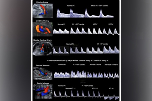
Fetal Doppler
A Fetal Doppler is a specialized ultrasound examination that evaluates blood flow between the mother, placenta, umbilical cord, and baby during pregnancy. By assessing fetal circulation, this advanced Doppler study helps determine whether the baby is receiving adequate oxygen and nutrients, making it an essential investigation for monitoring high-risk pregnancies and supporting healthy fetal development.
At Dr. Dere Sonography & X-Ray Centre, Fetal Doppler studies are performed by Dr. Prajakta Samir Dere, Consultant Radiologist with specialized expertise in Obstetric, Fetal & Gynecological Imaging. Using advanced Doppler ultrasound technology and internationally accepted fetal assessment protocols, every examination is performed with exceptional precision to provide accurate information regarding fetal well-being and placental circulation.
Unlike a routine pregnancy ultrasound, a Fetal Doppler focuses on evaluating blood flow through important fetal and maternal blood vessels. It helps detect signs of placental insufficiency, fetal growth restriction (FGR), fetal anemia, and other conditions that may affect your baby's growth and overall health. Early identification of these changes allows your obstetrician to plan timely treatment and appropriate pregnancy management.
Fetal Doppler is commonly recommended during the third trimester, especially in pregnancies complicated by hypertension, diabetes, fetal growth restriction, twin pregnancies, or other high-risk conditions. It provides valuable information that supports safer pregnancy care and helps determine the best timing and mode of delivery whenever required.
Conditions Evaluated During a Fetal Doppler Study
A Fetal Doppler study provides detailed information about fetal circulation and placental function, helping identify conditions that may affect your baby's growth and well-being.
- Assessment of umbilical artery blood flow.
- Evaluation of middle cerebral artery (MCA) blood flow.
- Assessment of uterine artery circulation.
- Evaluation of ductus venosus when clinically indicated.
- Placental blood circulation assessment.
- Fetal Growth Restriction (FGR) evaluation.
- Assessment of fetal oxygen supply.
- Monitoring of high-risk pregnancies.
- Evaluation of twin and multiple pregnancies.
- Monitoring pregnancies complicated by hypertension or diabetes.
- Assessment of suspected fetal anemia.
- Overall fetal well-being before delivery.
When is a Fetal Doppler Recommended?
Your obstetrician may recommend a Fetal Doppler examination whenever detailed assessment of fetal blood circulation is required. This investigation is particularly valuable for monitoring pregnancies where closer fetal surveillance is necessary.
- When fetal growth restriction (FGR) is suspected.
- In pregnancies complicated by hypertension or preeclampsia.
- For gestational diabetes requiring fetal monitoring.
- When reduced fetal movements are reported.
- For twin or multiple pregnancies.
- When placental insufficiency is suspected.
- Following an abnormal Growth Scan.
- To monitor pregnancies with previous obstetric complications.
- For pregnancies requiring serial fetal surveillance.
- Whenever advised by your obstetrician to evaluate fetal well-being.
Signs & Symptoms That May Require a Fetal Doppler Study
While a Fetal Doppler examination is commonly advised for high-risk pregnancies, your obstetrician may also recommend this scan whenever there is a concern about your baby's growth, placental function, or overall well-being. Early assessment of fetal blood circulation helps identify potential complications and allows timely medical intervention to support a healthy pregnancy.
- Reduced or decreased fetal movements.
- Suspected Fetal Growth Restriction (FGR).
- High blood pressure or preeclampsia during pregnancy.
- Gestational diabetes requiring fetal surveillance.
- Low amniotic fluid (Oligohydramnios).
- Abnormal findings on a previous Growth Scan.
- Twin or multiple pregnancy.
- History of previous pregnancy complications.
- Any pregnancy requiring close fetal monitoring.
What Does a Fetal Doppler Assess?
A Fetal Doppler study evaluates blood circulation through important maternal and fetal blood vessels, providing valuable information about oxygen delivery, placental function, and your baby's overall health. This examination helps your obstetrician identify early signs of fetal compromise and make informed decisions regarding pregnancy management.
Umbilical Artery Doppler
The umbilical artery carries blood between the placenta and your baby. Doppler assessment helps determine whether the placenta is supplying adequate oxygen and nutrients to support healthy fetal growth.
Middle Cerebral Artery (MCA) Doppler
The MCA Doppler evaluates blood flow to the baby's brain. It is particularly useful in detecting fetal anemia, monitoring fetal adaptation to reduced oxygen supply, and assessing overall fetal well-being.
Uterine Artery Doppler
This examination evaluates blood flow through the uterine arteries supplying the placenta. Abnormal findings may indicate an increased risk of preeclampsia, placental insufficiency, or fetal growth restriction.
Ductus Venosus Assessment
When clinically indicated, the ductus venosus is assessed to evaluate fetal cardiac function and blood circulation. This is particularly useful in selected high-risk pregnancies and specialized fetal assessments.
Placental Blood Circulation
The scan evaluates placental function by assessing blood flow between the mother and baby. Healthy placental circulation is essential for providing adequate oxygen and nutrition throughout pregnancy.
Fetal Oxygenation & Well-being
By combining Doppler measurements from different blood vessels, the examination helps determine whether your baby is receiving sufficient oxygen and whether additional monitoring or intervention may be required.
Monitoring High-Risk Pregnancies
Fetal Doppler plays an important role in monitoring pregnancies complicated by hypertension, diabetes, fetal growth restriction, multiple pregnancy, or previous obstetric complications, allowing your obstetrician to provide individualized pregnancy care.
How is a Fetal Doppler Performed?
A Fetal Doppler study is a safe, painless, and non-invasive ultrasound examination performed using advanced Doppler ultrasound technology. The procedure usually takes approximately 20 to 30 minutes, depending on the baby's position and the number of blood vessels that need to be assessed.
- Your pregnancy history and previous ultrasound reports are reviewed before the examination.
- A water-based ultrasound gel is gently applied over the abdomen.
- Advanced Doppler ultrasound is used to evaluate blood flow through the umbilical artery, uterine arteries, middle cerebral artery, and other vessels when required.
- Fetal heartbeat, fetal movements, placental circulation, and overall fetal well-being are simultaneously assessed.
- All Doppler findings are carefully analysed and compared with the baby's gestational age.
- A comprehensive Doppler report is prepared to assist your obstetrician in planning further pregnancy care.
Benefits of a Fetal Doppler Study
Fetal Doppler is an invaluable investigation for monitoring fetal health during pregnancy. It provides important information that helps detect complications early and supports safer pregnancy management.
- Evaluates blood circulation between the placenta and baby.
- Assesses fetal oxygen and nutrient supply.
- Detects placental insufficiency at an early stage.
- Monitors pregnancies affected by fetal growth restriction (FGR).
- Supports monitoring of high-risk pregnancies.
- Assists in determining the safest timing for delivery when required.
- Provides reassurance regarding your baby's overall well-being.
- Supports timely medical decisions for improved maternal and fetal outcomes.
Preparation Before a Fetal Doppler Study
A Fetal Doppler study requires minimal preparation and is performed in the same comfortable manner as a routine pregnancy ultrasound. Following a few simple instructions helps obtain clear Doppler waveforms and high-quality images for accurate assessment of fetal circulation.
- Carry your doctor's prescription along with all previous ultrasound and Doppler reports.
- A full bladder is generally not required unless specifically advised by your doctor.
- Wear comfortable, loose-fitting clothing to facilitate the examination.
- Reach the diagnostic centre 10–15 minutes before your scheduled appointment.
- Bring your antenatal records and recent laboratory investigations if available.
- Follow any additional instructions provided by your obstetrician or radiologist.
Is a Fetal Doppler Study Safe?
Yes. A Fetal Doppler study is a safe, painless, and non-invasive ultrasound examination that uses sound waves to evaluate blood flow within the placenta and fetal blood vessels. It does not use radiation and is widely recommended for monitoring pregnancies where detailed assessment of fetal circulation is required.
At Dr. Dere Sonography & X-Ray Centre, every Doppler examination is performed using advanced ultrasound technology while adhering to internationally accepted obstetric imaging protocols, ensuring reliable results, patient comfort, and the highest standards of maternal and fetal care.
Frequently Asked Questions
What is the difference between a Growth Scan and a Fetal Doppler?
A Growth Scan primarily evaluates your baby's size, growth, amniotic fluid, and placental appearance, whereas a Fetal Doppler specifically measures blood flow within important maternal and fetal blood vessels to assess oxygen and nutrient supply to the baby.
When is a Fetal Doppler usually performed?
It is commonly recommended during the third trimester, particularly in pregnancies affected by hypertension, diabetes, fetal growth restriction, twin pregnancy, reduced fetal movements, or suspected placental insufficiency.
Is a Fetal Doppler painful?
No. The examination is completely painless and similar to a routine pregnancy ultrasound. Most Doppler studies are completed within 20 to 30 minutes.
Will every pregnant woman need a Fetal Doppler?
Not always. While many women undergo routine pregnancy ultrasounds, a Fetal Doppler is usually advised when your obstetrician requires additional information regarding placental blood flow or fetal well-being.
Can a Fetal Doppler help prevent pregnancy complications?
A Fetal Doppler cannot prevent complications, but it helps identify reduced blood flow and fetal compromise at an early stage, allowing timely medical intervention and appropriate pregnancy management.
Why Choose Dr. Prajakta Samir Dere for a Fetal Doppler Study?
- Consultant Radiologist with specialized expertise in Obstetric, Fetal & Gynecological Imaging.
- Advanced assessment of fetal circulation using high-resolution Color Doppler ultrasound technology.
- Comprehensive evaluation of the Umbilical Artery, Middle Cerebral Artery (MCA), Uterine Arteries, and other fetal vessels whenever clinically indicated.
- Expert monitoring of high-risk pregnancies, fetal growth restriction, and placental insufficiency.
- Evidence-based reporting following internationally accepted fetal Doppler imaging guidelines.
- Accurate, timely, and clinically reliable reports to support your obstetrician in making informed pregnancy care decisions.
- Patient-friendly environment with compassionate care for every expecting mother.
- Trusted prenatal imaging services focused on maternal and fetal well-being.
Supporting Healthy Pregnancies Through Advanced Fetal Monitoring
A Fetal Doppler study provides valuable information about your baby's blood circulation, placental function, and overall well-being during pregnancy. Early detection of circulatory changes allows your obstetrician to closely monitor fetal health, plan timely interventions when required, and support the safest possible pregnancy outcome for both mother and baby.
Schedule your Fetal Doppler Study with Dr. Prajakta Samir Dere at Dr. Dere Sonography & X-Ray Centre for advanced fetal circulation assessment, expert pregnancy imaging, and compassionate prenatal care using state-of-the-art ultrasound technology.