
Fetal Echocardiography
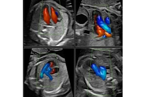
Fetal Echocardiography
Fetal Echocardiography is a specialized ultrasound examination performed during pregnancy to evaluate the structure and function of the baby's heart. It provides detailed assessment of fetal cardiac anatomy, heart chambers, valves, blood flow, and rhythm to identify possible congenital heart abnormalities at an early stage.
At Dr. Dere Sonography & X-Ray Centre, Fetal Echocardiography Screening is performed by Dr. Prajakta Samir Dere, Consultant Radiologist with expertise in Obstetric, Fetal & Gynecological Imaging. Using advanced ultrasound technology, detailed fetal cardiac evaluation is performed with precision while ensuring the comfort and safety of the mother and baby.
The fetal heart undergoes significant development during pregnancy, and specialized cardiac screening helps assess the baby's heart structures and function. This examination provides valuable information to obstetricians and fetal medicine specialists for appropriate pregnancy management.
Fetal Echocardiography is commonly recommended for pregnancies with increased risk of congenital heart disease or when abnormalities are suspected during routine ultrasound scans. Early detection allows better counselling, monitoring, and preparation for appropriate care after birth.
Conditions Evaluated During Fetal Echocardiography Screening
Fetal Echocardiography provides detailed evaluation of the developing fetal heart and helps identify various cardiac conditions during pregnancy.
- Congenital heart defects (CHD).
- Abnormal development of heart chambers.
- Heart valve abnormalities.
- Abnormal heart rhythm evaluation.
- Septal defects (holes between heart chambers).
- Abnormal blood flow patterns.
- Structural abnormalities of major blood vessels.
- Fetal cardiac function assessment.
- Follow-up evaluation of suspected heart abnormalities.
- Overall fetal cardiac well-being assessment.
When is Fetal Echocardiography Screening Recommended?
Your doctor may recommend Fetal Echocardiography when detailed evaluation of the baby's heart is required due to pregnancy risk factors, abnormal findings, or clinical indications.
- Abnormal findings on routine pregnancy ultrasound.
- Family history of congenital heart disease.
- Maternal diabetes during pregnancy.
- Pregnancy with increased risk of fetal abnormalities.
- Abnormal fetal heart rhythm detected on ultrasound.
- Suspected chromosomal or genetic conditions.
- Multiple pregnancy with clinical concerns.
- Whenever advised by your obstetrician or fetal medicine specialist.
Why is Fetal Echocardiography Screening Recommended?
Fetal Echocardiography Screening is recommended when detailed evaluation of the baby's heart structure and function is required during pregnancy. It helps identify possible congenital heart abnormalities early and supports appropriate pregnancy monitoring and delivery planning.
- Provides detailed evaluation of fetal heart anatomy.
- Helps detect congenital heart defects before birth.
- Assesses fetal heart chambers, valves, and blood flow.
- Evaluates fetal heart rhythm and function.
- Supports better pregnancy management in high-risk cases.
- Helps doctors plan appropriate care after delivery.
- Provides reassurance when fetal heart development appears normal.
- Allows early counselling and preparation for families when abnormalities are detected.
What Does Fetal Echocardiography Assess?
Fetal Echocardiography provides a detailed assessment of the developing baby's heart, including its structure, function, rhythm, and blood circulation patterns.
Heart Chambers Evaluation
The four chambers of the fetal heart are carefully examined to assess their size, structure, development, and overall function.
Heart Valve Assessment
The fetal heart valves are evaluated to check their movement, function, and blood flow patterns during the cardiac cycle.
Major Blood Vessel Evaluation
The major arteries and veins connected to the fetal heart are assessed to identify any structural or flow-related abnormalities.
Heart Rhythm Assessment
The fetal heart rate and rhythm are evaluated to identify irregular heartbeats or rhythm disturbances.
Cardiac Function Assessment
The pumping function of the fetal heart is evaluated to ensure proper cardiac performance and circulation.
Septal Defect Evaluation
The walls separating the heart chambers are assessed to identify possible defects or abnormalities.
Blood Flow Assessment
Doppler techniques are used to evaluate blood flow patterns through the fetal heart and major vessels.
Overall Fetal Cardiac Health
A comprehensive assessment of the fetal heart helps identify abnormalities and supports better prenatal care planning.
How is Fetal Echocardiography Screening Performed?
Fetal Echocardiography is a safe, non-invasive ultrasound examination performed using advanced imaging technology. A specialized ultrasound probe is gently moved over the mother's abdomen to obtain detailed images of the baby's heart. The examination usually takes around 30 to 60 minutes, depending on fetal position and the complexity of evaluation required.
- Your pregnancy history, previous ultrasound reports, and medical details are reviewed before the examination.
- The mother is positioned comfortably on the examination table.
- Ultrasound gel is applied over the abdomen for better image quality.
- Detailed images of the fetal heart are obtained using advanced ultrasound technology.
- Heart chambers, valves, rhythm, and blood flow are carefully evaluated.
- A detailed report is prepared for your obstetrician or fetal medicine specialist.
Benefits of Fetal Echocardiography Screening
Fetal Echocardiography provides important information about the baby's heart development and plays a valuable role in prenatal care, especially in high-risk pregnancies.
- Early detection of congenital heart abnormalities.
- Detailed assessment of fetal cardiac structures.
- Evaluation of fetal heart function and blood flow.
- Safe and radiation-free examination.
- Helps in planning appropriate pregnancy care.
- Provides valuable information for delivery planning.
- Supports early medical counselling when required.
- Improves confidence through detailed fetal heart evaluation.
Preparation Before Fetal Echocardiography Screening
Fetal Echocardiography Screening is a simple, safe, and comfortable ultrasound examination that requires minimal preparation. Following a few basic instructions helps obtain clear images of the baby's heart and ensures a smooth examination experience.
- Carry your doctor's prescription along with previous ultrasound reports and pregnancy records.
- Inform the radiologist about your pregnancy history, medical conditions, and previous scan findings.
- Wear comfortable clothing that allows easy access to the abdomen.
- Follow your doctor's instructions regarding food, water intake, or bladder preparation if advised.
- Bring any previous genetic screening or fetal scan reports if available.
- Arrive at the diagnostic centre 10 to 15 minutes before your scheduled appointment.
Is Fetal Echocardiography Screening Safe?
Yes. Fetal Echocardiography Screening is a completely safe, painless, and non-invasive ultrasound examination. It uses high-frequency sound waves to evaluate the baby's heart and does not involve radiation exposure. The procedure is considered safe for both mother and baby when performed as recommended by your doctor.
At Dr. Dere Sonography & X-Ray Centre, every Fetal Echocardiography Screening is performed using advanced ultrasound technology while maintaining strict standards of maternal comfort, fetal safety, privacy, and diagnostic accuracy.
Frequently Asked Questions
What is Fetal Echocardiography Screening?
Fetal Echocardiography Screening is a specialized ultrasound examination that evaluates the structure, function, rhythm, and blood flow of the baby's heart during pregnancy.
When is Fetal Echocardiography usually performed?
It is commonly performed during the second trimester of pregnancy, although the timing may vary depending on clinical requirements and your doctor's advice.
Is Fetal Echocardiography painful?
No. The examination is completely painless and similar to a routine ultrasound scan. A probe is gently moved over the mother's abdomen to capture detailed images of the fetal heart.
Why is Fetal Echocardiography advised during pregnancy?
It is advised when there is a need for detailed evaluation of the baby's heart due to risk factors, abnormal ultrasound findings, family history, or other medical indications.
Can Fetal Echocardiography detect all heart problems?
Fetal Echocardiography can detect many structural and functional heart abnormalities, but some conditions may only become apparent after birth or later stages of development.
How long does Fetal Echocardiography take?
The examination usually takes around 30 to 60 minutes, depending on fetal position, image quality, and the detailed assessment required.
Why Choose Dr. Prajakta Samir Dere for Fetal Echocardiography Screening?
- Consultant Radiologist with specialized expertise in Obstetric, Fetal & Gynecological Imaging.
- Advanced ultrasound technology for detailed fetal cardiac evaluation.
- Expert assessment of fetal heart chambers, valves, rhythm, and blood flow.
- Accurate prenatal diagnosis and comprehensive fetal heart screening.
- Compassionate and comfortable environment for expecting mothers.
- Detailed reporting to support obstetric and fetal medicine decisions.
- Strict safety standards followed during every ultrasound examination.
- Trusted fetal imaging services focused on mother and baby care.
Advanced Fetal Heart Screening for a Healthy Pregnancy
Fetal Echocardiography Screening plays an important role in prenatal care by providing detailed evaluation of the baby's heart development. Early identification of cardiac abnormalities helps doctors and families prepare for appropriate monitoring, delivery planning, and newborn care whenever required.
Schedule your Fetal Echocardiography Screening with Dr. Prajakta Samir Dere at Dr. Dere Sonography & X-Ray Centre for advanced fetal cardiac imaging, expert diagnosis, and compassionate pregnancy care using modern ultrasound technology.