
Transvaginal Ultrasound (TVS)
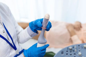
Transvaginal Ultrasound (TVS)
Transvaginal Ultrasound (TVS) is a highly accurate pelvic imaging examination that uses a specially designed ultrasound probe placed gently inside the vagina to obtain detailed images of the uterus, ovaries, cervix, endometrium, and surrounding pelvic structures. It plays a vital role in diagnosing gynecological conditions, infertility, early pregnancy complications, pelvic pain, abnormal uterine bleeding, and other reproductive health concerns.
At Dr. Dere Sonography & X-Ray Centre, Transvaginal Ultrasound examinations are performed by Dr. Prajakta Samir Dere, Consultant Radiologist with specialized expertise in Obstetric, Fetal & Gynecological Imaging. Using advanced high-resolution ultrasound technology, every examination provides exceptional image clarity, enabling accurate diagnosis while ensuring complete patient comfort, privacy, and confidentiality.
Unlike a routine abdominal ultrasound, a Transvaginal Ultrasound places the ultrasound probe closer to the pelvic organs, resulting in superior image quality and greater diagnostic accuracy. It is especially useful for evaluating the uterus, ovaries, endometrial lining, early pregnancy, infertility, ovarian cysts, fibroids, adenomyosis, endometriosis, and other pelvic abnormalities that may not be clearly visible on a transabdominal scan.
Whether you are experiencing pelvic pain, irregular menstrual cycles, abnormal vaginal bleeding, infertility, or require detailed evaluation during early pregnancy, a Transvaginal Ultrasound provides valuable diagnostic information that helps your gynecologist make informed treatment decisions while supporting your reproductive health.
Conditions Evaluated During Transvaginal Ultrasound
Transvaginal Ultrasound provides detailed visualization of the female reproductive organs, allowing accurate diagnosis and monitoring of numerous gynecological and early pregnancy conditions.
- Ovarian cysts and ovarian masses.
- Uterine fibroids.
- Adenomyosis.
- Polycystic Ovarian Syndrome (PCOS).
- Endometrial polyps and endometrial thickening.
- Abnormal uterine bleeding.
- Early pregnancy assessment.
- Ectopic pregnancy evaluation.
- Infertility assessment.
- Pelvic inflammatory disease (PID).
- Postmenopausal bleeding.
- Routine pelvic organ evaluation.
When is a Transvaginal Ultrasound Recommended?
Your gynecologist may recommend a Transvaginal Ultrasound whenever detailed pelvic imaging is required for diagnosis, treatment planning, fertility evaluation, or early pregnancy assessment.
- Irregular or abnormal menstrual bleeding.
- Pelvic or lower abdominal pain.
- Evaluation of infertility.
- Follicular monitoring during fertility treatment.
- Suspected ovarian cysts or uterine fibroids.
- Early pregnancy confirmation.
- Suspected ectopic pregnancy.
- Postmenopausal bleeding.
- Follow-up of previously diagnosed gynecological conditions.
- Whenever advised by your gynecologist for detailed pelvic evaluation.
Signs & Symptoms That May Require a Transvaginal Ultrasound
A Transvaginal Ultrasound (TVS) is recommended when a detailed examination of the female pelvic organs is required. It provides superior visualization of the uterus, ovaries, endometrium, and surrounding pelvic structures, helping your gynecologist accurately diagnose the underlying cause of your symptoms.
- Irregular, missed, or absent menstrual periods.
- Heavy or prolonged menstrual bleeding.
- Abnormal vaginal bleeding between periods.
- Persistent pelvic or lower abdominal pain.
- Pain during sexual intercourse.
- Difficulty conceiving or infertility evaluation.
- Suspected ovarian cysts, fibroids, or pelvic masses.
- Early pregnancy pain or vaginal spotting.
- Postmenopausal bleeding.
What Does a Transvaginal Ultrasound Assess?
Transvaginal Ultrasound provides highly detailed images of the female reproductive organs, making it one of the most accurate imaging techniques for diagnosing gynecological disorders, evaluating infertility, and assessing early pregnancy.
Uterus Assessment
The uterus is carefully evaluated for its size, shape, position, and internal structure to identify fibroids, adenomyosis, congenital uterine abnormalities, and other uterine conditions.
Endometrial Evaluation
The endometrial lining is examined for its thickness and appearance to detect polyps, hyperplasia, retained products, and other abnormalities associated with abnormal uterine bleeding or infertility.
Ovarian Assessment
Both ovaries are examined in detail to identify ovarian cysts, polycystic ovaries (PCOS), benign or complex ovarian masses, follicular development, and ovulation-related changes.
Cervical Assessment
The cervix is evaluated whenever clinically indicated to identify cervical abnormalities, cervical polyps, or structural changes requiring further gynecological assessment.
Early Pregnancy Evaluation
Transvaginal Ultrasound is highly effective for confirming an early intrauterine pregnancy, assessing fetal viability, detecting ectopic pregnancy, and evaluating the gestational sac, yolk sac, and embryo during the first trimester.
Pelvic Masses & Free Fluid
The examination helps detect pelvic masses, fluid collections, ruptured ovarian cysts, pelvic infections, and other abnormalities affecting the female pelvis.
Infertility Assessment
TVS plays a vital role in infertility evaluation by assessing follicular growth, ovarian reserve, endometrial development, ovulation, and pelvic anatomy during fertility treatment.
Overall Pelvic Health
All pelvic organs are systematically examined to provide a comprehensive assessment of reproductive health, allowing early diagnosis and effective treatment planning.
How is a Transvaginal Ultrasound Performed?
A Transvaginal Ultrasound is a safe, painless, and minimally invasive examination performed using a thin, specially designed ultrasound probe covered with a protective sterile sheath. The examination usually takes 15 to 30 minutes and provides highly detailed pelvic images that cannot always be obtained through an abdominal ultrasound.
- Your symptoms, medical history, and previous reports are reviewed before the examination.
- You will be asked to empty your bladder before the scan for optimal visualization.
- A sterile, lubricated ultrasound probe is gently inserted into the vagina with your consent.
- The uterus, endometrium, ovaries, cervix, and surrounding pelvic structures are carefully examined.
- Detailed measurements and diagnostic images are recorded.
- A comprehensive ultrasound report is prepared to assist your gynecologist in further treatment planning.
Benefits of a Transvaginal Ultrasound
Transvaginal Ultrasound offers superior image quality compared with routine pelvic ultrasound, making it one of the most valuable diagnostic tools for women's reproductive health.
- Provides exceptionally detailed images of pelvic organs.
- Early detection of ovarian cysts, fibroids, and pelvic abnormalities.
- Highly accurate assessment of the endometrium and uterus.
- Essential investigation for infertility evaluation and follicular monitoring.
- Excellent imaging for early pregnancy and ectopic pregnancy assessment.
- Supports timely diagnosis and individualized treatment planning.
- Safe, radiation-free, and well tolerated by most patients.
- Improves diagnostic accuracy for a wide range of gynecological conditions.
Preparation Before a Transvaginal Ultrasound
A Transvaginal Ultrasound (TVS) requires very little preparation and is performed in a comfortable, private, and hygienic environment. Unlike an abdominal pelvic ultrasound, a full bladder is generally not required. Following a few simple instructions helps ensure optimal image quality and a smooth examination.
- Carry your doctor's prescription, previous ultrasound reports, and relevant medical records.
- Empty your bladder before the examination unless instructed otherwise.
- Wear comfortable, loose-fitting clothing for convenience during the scan.
- Arrive 10 to 15 minutes before your scheduled appointment.
- Inform your radiologist about pregnancy, recent surgeries, infections, or any significant medical history.
- The procedure is performed only with your informed consent while maintaining complete privacy and dignity.
Is a Transvaginal Ultrasound Safe?
Yes. A Transvaginal Ultrasound is a safe, painless, and radiation-free imaging examination that uses high-frequency sound waves to produce detailed images of the pelvic organs. It is routinely performed worldwide for gynecological evaluation, infertility assessment, and early pregnancy imaging without exposing patients to radiation.
At Dr. Dere Sonography & X-Ray Centre, every Transvaginal Ultrasound is performed using advanced ultrasound technology with strict attention to patient comfort, privacy, hygiene, and internationally accepted imaging standards.
Frequently Asked Questions
Is a Transvaginal Ultrasound painful?
Most women experience little or no pain during the examination. You may feel mild pressure while the ultrasound probe is gently inserted, but the procedure is generally well tolerated and lasts only a few minutes.
Do I need a full bladder for a TVS?
No. Unlike a Transabdominal Ultrasound, a Transvaginal Ultrasound is usually performed with an empty bladder to provide clearer visualization of the pelvic organs.
Can a Transvaginal Ultrasound detect early pregnancy?
Yes. TVS is one of the most accurate imaging techniques for confirming an early intrauterine pregnancy, assessing fetal viability, and detecting ectopic pregnancy during the first trimester.
Is TVS useful for infertility evaluation?
Yes. It plays a key role in infertility investigations by assessing the uterus, ovaries, endometrial lining, follicular growth, ovulation, and other factors affecting fertility.
How long does the examination take?
Most Transvaginal Ultrasound examinations are completed within 15 to 30 minutes, depending on the clinical indication and the level of assessment required.
Why Choose Dr. Prajakta Samir Dere for Transvaginal Ultrasound?
- Consultant Radiologist with specialized expertise in Obstetric, Fetal & Gynecological Imaging.
- Advanced high-resolution Transvaginal Ultrasound (TVS) for precise pelvic imaging.
- Expert evaluation of the uterus, ovaries, endometrium, cervix, and surrounding pelvic structures.
- Accurate diagnosis of fibroids, ovarian cysts, PCOS, infertility-related conditions, early pregnancy concerns, and abnormal uterine bleeding.
- Evidence-based reporting using modern ultrasound technology for reliable clinical decision-making.
- Compassionate, confidential, and patient-centred care with complete respect for privacy and comfort.
- Prompt, comprehensive reports to assist your gynecologist in planning the most appropriate treatment.
- Trusted women's imaging services focused on early diagnosis and lifelong reproductive health.
Advanced Pelvic Imaging for Accurate Women's Healthcare
Timely diagnosis plays an important role in maintaining women's reproductive health. Whether you require evaluation for pelvic pain, abnormal bleeding, infertility, ovarian cysts, fibroids, or early pregnancy, a Transvaginal Ultrasound provides highly detailed images that help ensure accurate diagnosis and effective treatment planning.
Schedule your Transvaginal Ultrasound (TVS) with Dr. Prajakta Samir Dere at Dr. Dere Sonography & X-Ray Centre for advanced pelvic imaging, expert diagnosis, and compassionate women's healthcare using state-of-the-art ultrasound technology.