
Portal Doppler
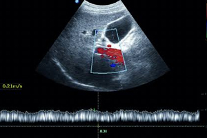
Portal Doppler
Portal Doppler is an advanced, safe, and non-invasive ultrasound examination that evaluates blood flow within the portal vein and the major blood vessels supplying the liver. It helps diagnose portal hypertension, portal vein thrombosis, liver cirrhosis-related vascular changes, and other hepatic vascular disorders while providing accurate real-time assessment without radiation exposure.
At Dr. Dere Sonography & X-Ray Centre, Portal Doppler examinations are performed by Dr. Sameer Dere, Consultant Radiologist with extensive expertise in Vascular and Diagnostic Ultrasound Imaging. Using advanced Color Doppler technology, every examination provides detailed evaluation of portal venous circulation, hepatic blood flow, and liver vascular health with excellent diagnostic accuracy.
Portal Doppler combines conventional ultrasound with Color Doppler imaging to assess both the liver anatomy and blood flow through the portal vein, hepatic artery, and hepatic veins. It is widely used for evaluating chronic liver disease, portal hypertension, liver cirrhosis, portal vein thrombosis, and post-liver transplant vascular assessment.
Whether you have chronic liver disease, unexplained abdominal swelling, suspected portal hypertension, abnormal liver function tests, or require follow-up after liver surgery or transplantation, Portal Doppler provides valuable diagnostic information that supports accurate diagnosis and timely treatment planning.
Conditions Evaluated During Portal Doppler
Portal Doppler provides comprehensive assessment of the portal venous system and hepatic circulation, helping diagnose a wide range of liver-related vascular disorders.
- Portal hypertension.
- Portal vein thrombosis.
- Liver cirrhosis with vascular assessment.
- Hepatic vein abnormalities.
- Portal vein obstruction.
- Budd-Chiari Syndrome.
- Assessment after liver transplantation.
- Evaluation of liver vascular tumors.
- Monitoring chronic liver disease.
- Post-operative hepatic vascular follow-up.
- Assessment of abnormal liver blood flow.
- Routine hepatic vascular evaluation.
When is a Portal Doppler Recommended?
Your doctor may recommend a Portal Doppler whenever detailed evaluation of portal venous circulation or liver blood flow is required to diagnose liver disease, vascular abnormalities, or monitor treatment outcomes.
- Known liver cirrhosis or chronic liver disease.
- Suspected portal hypertension.
- Abdominal swelling (Ascites).
- Suspected portal vein thrombosis.
- Abnormal liver function tests.
- Follow-up after liver transplantation or surgery.
- Evaluation of hepatic vascular disorders.
- Monitoring progression of chronic liver disease.
- Routine assessment whenever advised by your gastroenterologist, hepatologist, or physician.
Signs & Symptoms That May Require a Portal Doppler
A Portal Doppler examination is recommended whenever symptoms or clinical findings suggest abnormalities in liver blood circulation or the portal venous system. It helps assess blood flow to and from the liver, detect vascular complications, and support early diagnosis of chronic liver diseases.
- Known liver cirrhosis or chronic liver disease.
- Persistent abdominal swelling (Ascites).
- Suspected portal hypertension.
- Abnormal liver function tests.
- Suspected portal vein thrombosis.
- Enlarged spleen (Splenomegaly).
- Follow-up after liver transplantation or hepatic surgery.
- Monitoring chronic liver disease and portal circulation.
- Routine evaluation whenever advised by your gastroenterologist, hepatologist, or physician.
What Does a Portal Doppler Assess?
Portal Doppler provides a detailed evaluation of the portal venous system and hepatic blood circulation. It measures blood flow direction, velocity, and vessel patency while helping diagnose vascular disorders affecting the liver and surrounding abdominal organs.
Portal Vein Assessment
The portal vein is carefully evaluated for its size, blood flow direction, flow velocity, thrombosis, obstruction, and changes associated with portal hypertension or chronic liver disease.
Hepatic Vein Evaluation
The hepatic veins are examined to assess venous drainage from the liver and detect abnormalities such as Budd-Chiari Syndrome, venous obstruction, or impaired hepatic circulation.
Hepatic Artery Assessment
The hepatic artery is evaluated for blood flow characteristics, vascular narrowing, post-transplant complications, and adequate arterial supply to the liver.
Portal Hypertension Evaluation
Portal Doppler helps detect portal hypertension by assessing portal vein diameter, blood flow patterns, collateral circulation, and other vascular changes associated with advanced liver disease.
Portal Vein Thrombosis
The examination accurately identifies blood clots within the portal vein, evaluates the extent of thrombosis, and assesses its impact on liver circulation.
Post-Liver Transplant Assessment
Portal Doppler plays an important role after liver transplantation by evaluating portal venous flow, hepatic artery patency, hepatic vein drainage, and early detection of vascular complications.
Chronic Liver Disease Monitoring
Patients with cirrhosis and chronic liver disease benefit from regular Portal Doppler examinations to monitor progression of vascular changes and evaluate response to treatment.
Overall Hepatic Vascular Health
The examination provides a comprehensive assessment of liver blood circulation, helping your doctor diagnose vascular disorders accurately and plan the most appropriate management.
How is a Portal Doppler Performed?
Portal Doppler is a safe, painless, and non-invasive imaging examination performed using advanced Color Doppler Ultrasound technology. The procedure generally takes between 30 and 45 minutes and provides detailed real-time evaluation of portal venous and hepatic blood flow without radiation exposure.
- Your symptoms, medical history, and previous imaging reports are reviewed before the examination.
- You will usually be advised to fast for 6 to 8 hours before the examination.
- A water-based ultrasound gel is applied over the upper abdomen.
- The ultrasound probe is gently moved across the abdomen to evaluate the liver and portal blood vessels.
- Color Doppler imaging records blood flow direction, velocity, and vascular waveforms.
- A comprehensive Doppler report is prepared to assist your treating specialist in further management.
Benefits of a Portal Doppler
Portal Doppler is one of the most reliable investigations for evaluating liver blood circulation because it provides accurate, real-time vascular assessment while remaining completely safe and radiation-free.
- Detailed evaluation of portal vein and hepatic circulation.
- Early diagnosis of portal hypertension and portal vein thrombosis.
- Accurate assessment of hepatic artery and hepatic vein blood flow.
- Monitoring chronic liver disease and cirrhosis.
- Essential evaluation after liver transplantation and hepatic surgery.
- Safe, painless, and completely radiation-free examination.
- Supports early diagnosis and individualized treatment planning.
- Provides comprehensive assessment of liver vascular health.
Preparation Before a Portal Doppler
Portal Doppler is a safe and non-invasive imaging examination that requires a few simple preparations to obtain clear visualization of the liver and portal venous system. Following the recommended instructions helps improve image quality and ensures a more accurate assessment of hepatic blood circulation.
- Carry your doctor's prescription along with previous ultrasound, Doppler, CT/MRI scans, and relevant medical records.
- Fast for 6 to 8 hours before the examination unless your doctor advises otherwise.
- You may take regular medications with small sips of water if permitted by your doctor.
- Wear loose and comfortable clothing for easy abdominal examination.
- Arrive at the diagnostic centre 10 to 15 minutes before your scheduled appointment.
- Inform your radiologist about any previous liver surgery, liver transplantation, vascular procedures, or ongoing treatment for liver disease.
Is a Portal Doppler Safe?
Yes. Portal Doppler is a completely safe, painless, and radiation-free imaging examination that uses high-frequency sound waves to evaluate blood flow within the portal vein and hepatic blood vessels. It can be safely repeated whenever follow-up evaluation of liver circulation is required.
At Dr. Dere Sonography & X-Ray Centre, every Portal Doppler examination is performed using advanced Color Doppler technology while maintaining the highest standards of patient comfort, diagnostic accuracy, hygiene, and evidence-based reporting.
Frequently Asked Questions
Why is fasting required before a Portal Doppler?
Fasting helps reduce bowel gas, allowing clearer visualization of the liver, portal vein, and surrounding abdominal blood vessels. This improves the accuracy of the examination and the quality of diagnostic images.
What conditions can a Portal Doppler detect?
Portal Doppler helps diagnose portal hypertension, portal vein thrombosis, Budd-Chiari Syndrome, hepatic vascular abnormalities, chronic liver disease, cirrhosis-related vascular changes, and complications following liver transplantation.
Is the examination painful?
No. The examination is completely painless. A water-based gel is applied over the upper abdomen, and the ultrasound probe is gently moved across the skin to evaluate blood flow within the liver vessels.
How long does a Portal Doppler take?
Most Portal Doppler examinations are completed within 30 to 45 minutes, depending on the complexity of the vascular assessment and the patient's condition.
Can Portal Doppler be repeated for follow-up?
Yes. Since it does not involve radiation, Portal Doppler can be safely repeated to monitor chronic liver disease, evaluate treatment response, or assess blood flow after liver surgery or transplantation.
Why Choose Dr. Sameer Dere for Portal Doppler?
- Consultant Radiologist with extensive expertise in Vascular and Hepatobiliary Ultrasound Imaging.
- Advanced Color Doppler technology for accurate evaluation of portal venous and hepatic circulation.
- Expert assessment of portal hypertension, portal vein thrombosis, liver cirrhosis, and hepatic vascular disorders.
- Comprehensive evaluation before and after liver transplantation and hepatic surgery.
- Evidence-based reporting with precise findings for effective clinical decision-making.
- Comfortable, patient-friendly, and hygienic diagnostic environment.
- Prompt reporting for faster consultation with physicians, gastroenterologists, hepatologists, and liver surgeons.
- Trusted diagnostic imaging services focused on early diagnosis, patient safety, and long-term liver health.
Advanced Portal Venous Imaging for Better Liver Health
Healthy blood flow through the portal venous system is essential for proper liver function. Early diagnosis of portal venous and hepatic vascular disorders helps prevent serious complications associated with chronic liver disease and portal hypertension. Portal Doppler provides detailed real-time evaluation of liver blood circulation, supporting accurate diagnosis, timely treatment, and long-term monitoring.
Schedule your Portal Doppler with Dr. Sameer Dere at Dr. Dere Sonography & X-Ray Centre for advanced hepatic vascular imaging, expert diagnosis, and compassionate patient care using state-of-the-art Color Doppler technology.